
A cardiac assessment in nursing is a structured evaluation of the heart and peripheral circulation to identify early signs of cardiovascular compromise. The core components are history, inspection, palpation, auscultation, and peripheral vascular assessment. Below is a clear, clinically useful breakdown grounded in current nursing references.
🫀 Essential Components of a Nursing Cardiac Assessment
Takeaway: A complete cardiac assessment includes focused history, vital signs, inspection, palpation, auscultation, and peripheral vascular evaluation, each revealing different aspects of cardiac function. Lippincott NursingCenter+1
📝 1. Focused Cardiac History
- Chest pain: onset, duration, character, radiation, aggravating/relieving factors.
- Dyspnea or orthopnea.
- Palpitations or irregular heartbeat.
- Edema or sudden weight gain.
- Syncope or dizziness.
- Past history: hypertension, hyperlipidemia, diabetes, CAD, valve disease, rheumatic fever.
- Medications: beta‑blockers, antihypertensives, anticoagulants. nurseslab.in
👀 2. Inspection
- Skin color: pallor, cyanosis.
- Respiratory effort and work of breathing.
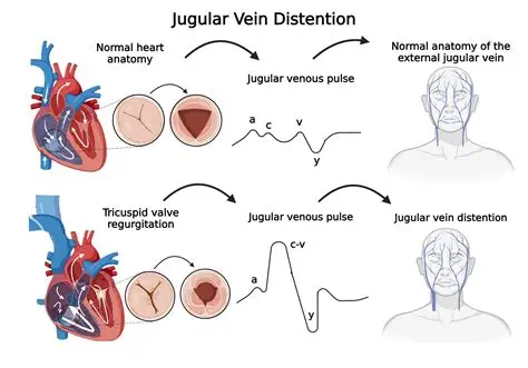
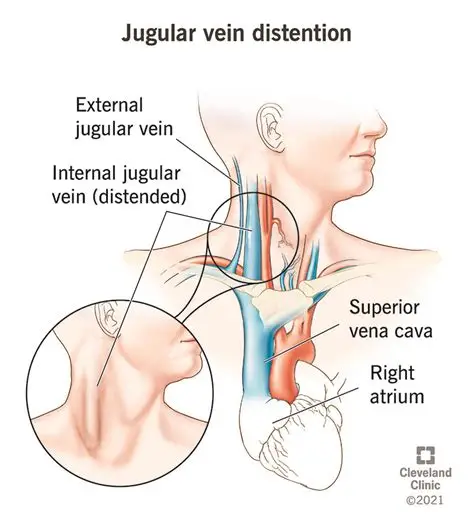
- Jugular venous distention (JVD).
- Chest wall for heaves or visible pulsations.
- Extremities for edema, temperature, capillary refill. Lippincott NursingCenter
✋ 3. Palpation
- Carotid pulses (one at a time): amplitude, symmetry.
- Precordium: PMI at 5th ICS, mid‑clavicular line.
- Peripheral pulses: radial, dorsalis pedis, posterior tibial.
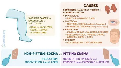
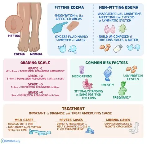
- Edema: pitting vs non‑pitting. Montgomery College Pressbooks Network
🎧 4. Auscultation
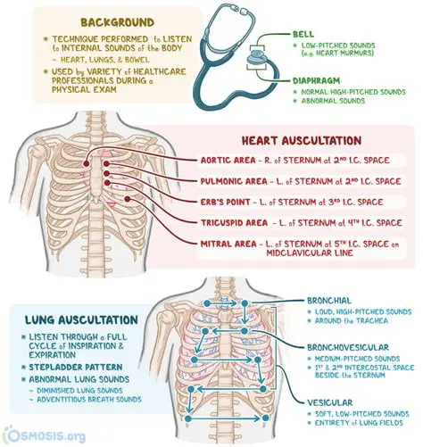
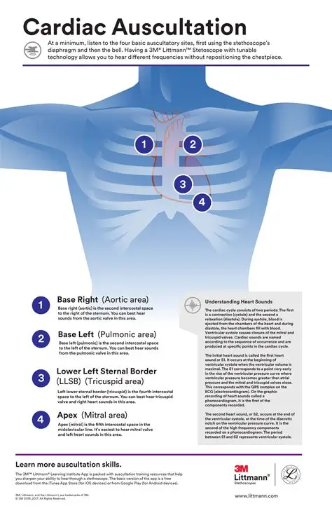
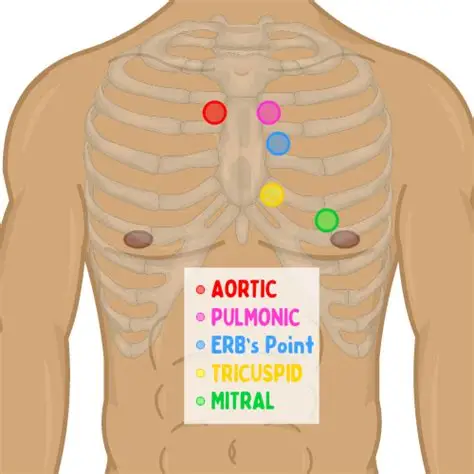
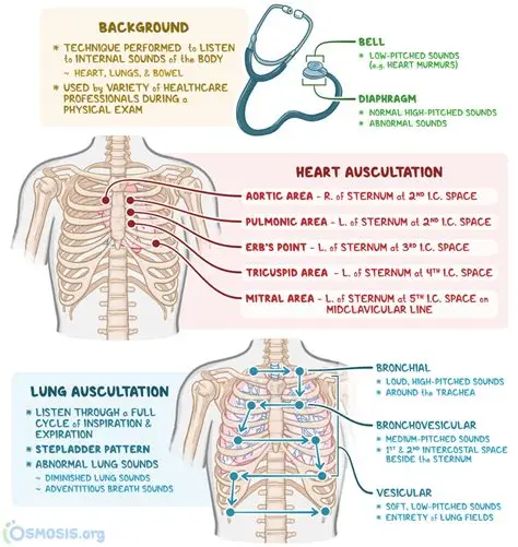
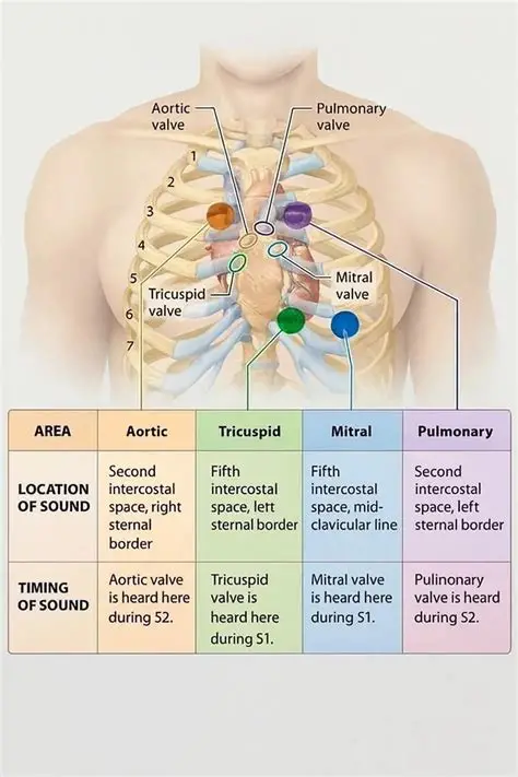
Auscultate with diaphragm and bell at the five cardiac landmarks:
- Aortic – 2nd ICS, RSB
- Pulmonic – 2nd ICS, LSB
- Erb’s point – 3rd ICS, LSB
- Tricuspid – 4th ICS, LSB
- Mitral (apex) – 5th ICS, MCL
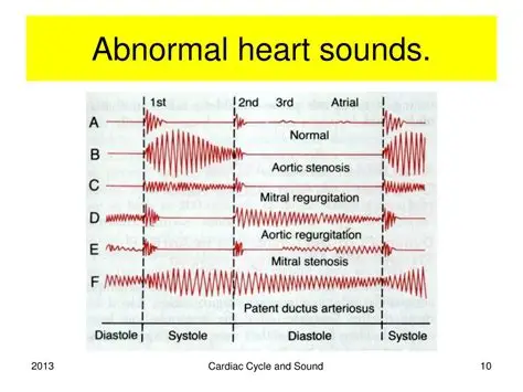
Listen for:
- S1, S2 (normal)
- S3, S4, murmurs, rubs, clicks Lecturio

🦵 5. Peripheral Vascular Assessment
- Color, temperature, moisture.
- Capillary refill (<2 seconds).
- Peripheral pulses: rate, rhythm, amplitude.
- Edema grading (1+ to 4+). Montgomery College Pressbooks Network
📋 6. Vital Signs
- Blood pressure (compare both arms if abnormal).
- Heart rate and rhythm.
- Respiratory rate.
- SpO₂. Lippincott NursingCenter
🧠 Putting It All Together: What Nurses Look For
- Signs of poor perfusion: cool extremities, delayed cap refill, weak pulses.
- Signs of fluid overload: JVD, edema, crackles, weight gain.
- Signs of cardiac ischemia: chest pain, diaphoresis, pallor.
- Signs of arrhythmia: irregular pulse, palpitations, dizziness.



🫀 Full Cardiac Assessment Note (Detailed Example)
Chief Complaint: Patient presents with intermittent chest pressure and shortness of breath on exertion.
Subjective:
Patient reports onset of substernal chest pressure approximately 3 hours prior to arrival. Describes sensation as “tight and heavy,” non‑radiating, lasting 5–10 minutes at a time. Symptoms worsen with walking and improve with rest. Patient reports associated shortness of breath and mild nausea during episodes. Denies diaphoresis, palpitations, dizziness, syncope, or recent illness. No history of trauma.
Past medical history includes hypertension and hyperlipidemia. Patient takes lisinopril 10 mg daily and atorvastatin 20 mg nightly. Denies missed doses. No known drug allergies.
Pain rated 5/10 during episodes, currently 2/10 at rest.
Objective:
General: Patient alert and oriented ×4, speaking in full sentences, appears mildly anxious but in no acute distress.
Vital Signs:
- BP: 148/88 mmHg
- HR: 96 bpm, regular
- RR: 20/min, unlabored
- SpO₂: 97% on room air
- Temp: 36.8°C
Inspection: Skin warm, slightly pale. No cyanosis. No visible chest wall deformities. No jugular venous distention noted at 45°. No peripheral edema.
Palpation:
- PMI palpable at 5th ICS, MCL, non‑displaced.
- No heaves or thrills.
- Peripheral pulses 2+ and equal bilaterally (radial, dorsalis pedis, posterior tibial).
- No lower‑extremity edema; capillary refill <2 seconds.
Auscultation:
- S1 and S2 present, regular.
- No murmurs, rubs, or gallops appreciated.
- No S3 or S4.
Respiratory: Lungs clear to auscultation bilaterally; no crackles or wheezes.
Assessment:
Patient with intermittent exertional chest pressure and dyspnea. Hemodynamically stable. Cardiac exam unremarkable aside from elevated BP. No signs of acute heart failure or peripheral hypoperfusion. Symptoms concerning for possible angina; requires further evaluation.
Plan:
- Continuous cardiac monitoring initiated.
- 12‑lead ECG obtained; results pending provider review.
- Vital signs q15 minutes.
- Patient instructed to remain resting and report any recurrence of chest pressure, shortness of breath, or new symptoms.
- Provider notified for immediate evaluation.
- Prepare for potential labs (troponin, CBC, CMP) and chest X‑ray per provider orders.
I


https://youtu.be/o802ujSSGO0?si=Eg6CYrAsNLq_ZrZ7
https://youtu.be/scP_76hXXyg?si=q2X0Rk7Rm076aWPh